Lameness
Lameness is a term used to describe a horse's gait being negatively affected by pain or a restriction in the typical range of movement, and it is a common problem in both pleasure and competition horses. Whilst in some horses the cause of the lameness may be easily determined – in others, a more detailed lameness work up is required. The severity of lameness is measured on a gradient scale, and in some horses lameness may be very subtle and difficult to recognise and only reveal itself as a loss of performance or difficulty or resistance in performing some activities.
At Agnew Equine, all of our vets are experienced at diagnosing and treating lameness cases. In addition, Stuart is one of only 21 vets in the country who is an Advanced Practitioner in Equine Orthopaedics from the Royal College of Veterinary Surgeons, so he is uniquely qualified to work with and assess any tricky lameness issues at your own stables – with no need to refer to a hospital for assessment.
We go through a number of steps to help determine the cause of your horse or pony’s lameness. To start, the history of the horse is important. What are the presenting signs, how long has the horse been lame, is it persistent or intermittent lameness, when do you notice the lameness, does the horse warm up and improve? All these will aid us when trying to figure out the cause. Then we would go through the following steps, until we are satisfied we’ve found the source of the lameness:
1) Physical examination
A careful examination of the horse is necessary both visually and with manual palpation. This is to identify any abnormalities such as asymmetry or limb swelling which could suggest the cause of the lameness. As a common culprit of lameness, the foot requires special attention and hoof testers are routinely used.
2) Walk and trot up in a straight line
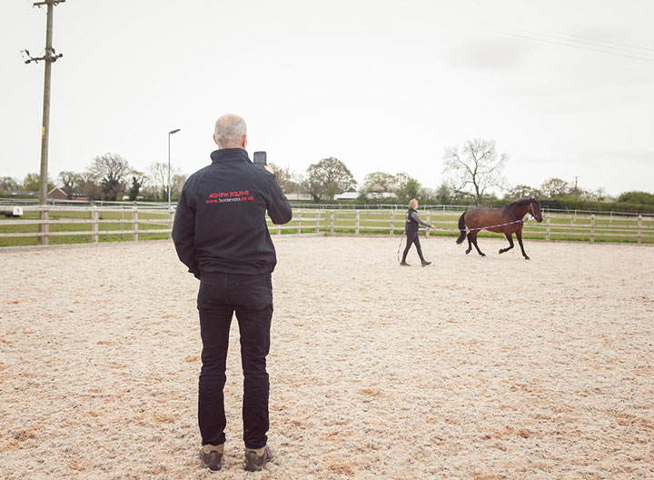
The majority of lamenesses will be assessed further by walking and trotting in a straight line ideally on a hard, level surface. This will help to identify the leg (or legs) where the horse is lame. We also evaluate the gait of the horse – as the way in which the horse moves and loads the limb can sometimes give clues to the site of pain and cause of the lameness. Our Equinosis® Lameness Locator® can be used to help identify the specific area of lameness – for more information on how this works click here.
3) Flexion tests
Flexion tests involve holding the leg (or the section of the leg) up before trotting the horse away. Flexion tests increase pressure on joints and surrounding structures and may exacerbate lameness associated with the area that has been stressed. The response or lack of a response will all add information when determining the site and causation of a lameness.
4) Lunging / ridden exercise
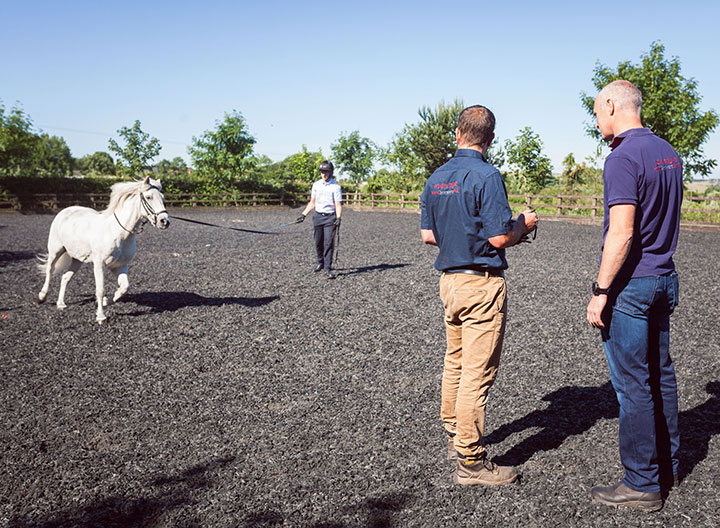
Lunging the horse on a surface is of great importance when dealing with troublesome lameness issues. The character and nature of the lameness will often appear as a certain pattern that can be identified when lunged. For example, a ligament or tendon strain may show lameness more obviously when the leg is on the outside of a circle. On occasion, ridden assessment will be useful.
5) Nerve blocks
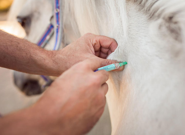
Nerve blocks (or diagnostic regional analgesia) uses local anaesthetic to ‘numb’ along the nerves a specific area of a horse’s leg. If the painful area is nerve blocked and the lameness improves – then it can be inferred that the site of the pain causing the lameness has been identified. Agnew Equine vets routinely perform nerve and joint blocks efficiently and safely at our client’s home premises. If the case requires it 2 vets will attend to perform the nerve block (all at no extra cost!)
6) Joint blocks
A joint block is performed under sterile, aseptic conditions. The area is cleaned and the joint or synovial structure is infiltrated with local anaesthetic. The lameness will then be assessed at various time points (5 mins, 10 minutes, 20 minutes) after injection. These tend to be more specific than nerve blocks, and are often used to further identify the structure affected.
Both nerve and joint blocks can be performed by our veterinary team on site at your stables.
7) Diagnostic Imaging
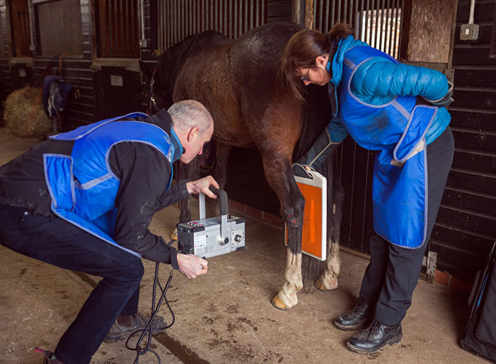
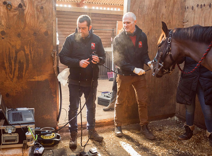
Once the site of the pain has been established, we use our mobile digital x-ray and/or ultrasound to determine a diagnosis. These are hospital-grade imaging modalities and are delivered stable-side. Occasionally, circumstances will require specialist imaging techniques. An MRI (magnetic resonance imaging) is particularly useful for investigating horses with foot pain and produces highly detailed images of the complicated soft tissue and bony structures of this area. Nuclear scintigraphy (bone scan) can be a useful tool in lameness and poor performance investigations with areas of damaged or inflamed bone identified as ‘hot spots’.
8) Treatment
Once the cause of the lameness has been established, our vets will advise you on appropriate treatments and management strategies. Broadly speaking, treatments can be subdivided into medical therapies such as anti-inflammatory medicines, regenerative treatments or surgery, such as arthroscopy. Once the treatment plan has been actioned, success rates will be maximised by using corrective shoeing, physiotherapy, rehabilitation and controlled exercise programs.
With the experience we have at Agnew Equine, we are able to diagnose and treat most cases of lameness ourselves. But sometimes, specialist input is required especially if an MRI, nuclear scintigraphy or surgery is required. As an independent practice, we are free and able to refer you to the best specialist for your particular case and we work alongside the referral hospital as well as with the follow-up care.

